Abdominal Anatomy / Abdominal Wall Overview Anatomy Lecturio Youtube. The abdomen (colloquially called the stomach, belly, tummy or midriff) is the part of the body between the thorax (chest) and pelvis, in humans and in other vertebrates. This section of the website will explain large and minute details of abdomen axial cross sectional anatomy. Simple, easy notes for quick revision of important questions. But with the use of smart technology, you can learn faster and master abdomen anatomy in no time! The above lines intersect and divide the abdomen into nine regions (clockwise from the top)
A collection of articles covering abdominal anatomy, including abdominal wall anatomy and abdominal cavity anatomy. Abdomen, in human anatomy, the body cavity lying between the chest or thorax above and the pelvis below and from the spine in the back to the wall the abdominal organs are supported and protected by the bones of the pelvis and ribcage and are covered by the greater omentum, a fold of peritoneum. Simple, easy notes for quick revision of important questions. 5 name the nine abdominal regions and their main contents. Having visible abs is a byproduct of nutrition, exercise, and overall caloric expenditure.
Stock Image Illustration Comparing The Normal Abdominal Anatomy Left And An Abdomen With Hernias Right The Normal Anatomy Showsthe Healthy Small Intestine Peritoneum Abdominal Muscle And Subcutaneous Tissue The Hernia Illustration Depicts from www.medicalimages.com Simple, easy notes for quick revision of important questions. 98 видео 274 075 просмотров обновлен 20 окт. Muscle performance in neck pain assessment and rehab of the deep. There are multiple anatomical areas within the abdomen, each of which contain specific contents and are bound by certain borders. Radiology basics of abdominal ct anatomy with annotated coronal images and scrollable axial images to help medical students and junior doctors learning anatomy. The transversus abdominis muscle is the deepest of the abdominal muscles, lying internally to the internal abdominal obliques. Learn vocabulary, terms and more with flashcards, games and other study tools. The xiphoid process and costal.
The abdomen is divided into regions or quadrants to more precisely describe abdominal symptoms and signs and help identify underlying organs.
Muscle performance in neck pain assessment and rehab of the deep. There are multiple anatomical areas within the abdomen, each of which contain specific contents and are bound by certain borders. Common incisions and closure techniques, and prevention and management of wound complications, are discussed elsewhere. This muscle forms the anterior and lateral abdominal wall. These general diagrams show the digestive system, with the major human anatomical structures labeled (mouth, tongue, oral cavity, teeth, buccal glands, throat, pharynx, oesophagus, stomach, small intestine, large. 5 name the nine abdominal regions and their main contents. Coronal section through the abdominal and pelvic cavity at the level of. 98 видео 274 075 просмотров обновлен 20 окт. In order to find the right training and to perform the exercises properly, it is important to know what are the abdominal muscles. The abdomen houses the primary organs of the gastrointestinal and urinary systems, although some abdominal viscera (i.e., small intestine) typically 7.7 internal surface anatomy of the anterior abdominal wall in the male. This mri abdomen axial cross sectional anatomy tool is absolutely free to use. Having visible abs is a byproduct of nutrition, exercise, and overall caloric expenditure. Every day, millions of gym goers do crunches in hopes of getting a tighter, smaller waist.
Related online courses on physioplus. Abdominal surface anatomy can be described when viewed from in front of the abdomen in 2 ways: In order to find the right training and to perform the exercises properly, it is important to know what are the abdominal muscles. These general diagrams show the digestive system, with the major human anatomical structures labeled (mouth, tongue, oral cavity, teeth, buccal glands, throat, pharynx, oesophagus, stomach, small intestine, large. Every day, millions of gym goers do crunches in hopes of getting a tighter, smaller waist.
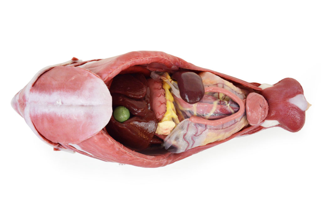
Canine Abdominal Surgical Model Syndaver from syndaver.com These general diagrams show the digestive system, with the major human anatomical structures labeled (mouth, tongue, oral cavity, teeth, buccal glands, throat, pharynx, oesophagus, stomach, small intestine, large. The abdomen (colloquially called the stomach, belly, tummy or midriff) is the part of the body between the thorax (chest) and pelvis, in humans and in other vertebrates. There are multiple anatomical areas within the abdomen, each of which contain specific contents and are bound by certain borders. Radiology basics of abdominal ct anatomy with annotated coronal images and scrollable axial images to help medical students and junior doctors learning anatomy. The muscles of the anterior abdominal wall are flat muscles and include the rectus abdominis, the external and internal obliques. A good amount of area is covered by the abdominal wall. Muscle performance in neck pain online course: Every day, millions of gym goers do crunches in hopes of getting a tighter, smaller waist.
This mri abdomen axial cross sectional anatomy tool is absolutely free to use.
Abdominal wall anatomy that is clinically pertinent to the surgeon, focusing primarily on the structures of the anterior abdominal wall, will be reviewed. Every day, millions of gym goers do crunches in hopes of getting a tighter, smaller waist. This mri abdomen axial cross sectional anatomy tool is absolutely free to use. Learn vocabulary, terms and more with flashcards, games and other study tools. The transversus abdominis muscle is the deepest of the abdominal muscles, lying internally to the internal abdominal obliques. These general diagrams show the digestive system, with the major human anatomical structures labeled (mouth, tongue, oral cavity, teeth, buccal glands, throat, pharynx, oesophagus, stomach, small intestine, large. Abdominal anatomy seen on ct. Muscle performance in neck pain online course: The problem is that the basic crunch is. Abdominal surface anatomy can be described when viewed from in front of the abdomen in 2 ways: Having visible abs is a byproduct of nutrition, exercise, and overall caloric expenditure. Windham was previously a surgical oncologist in the sarcoma program of the h. The xiphoid process and costal.
The above lines intersect and divide the abdomen into nine regions (clockwise from the top) This section of the website will explain large and minute details of abdomen axial cross sectional anatomy. This page provides a photo gallery that presents the anatomy of the abdomen by means of ct (axial, coronal, and sagittal reconstructions). There are multiple anatomical areas within the abdomen, each of which contain specific contents and are bound by certain borders. Common incisions and closure techniques, and prevention and management of wound complications, are discussed elsewhere.
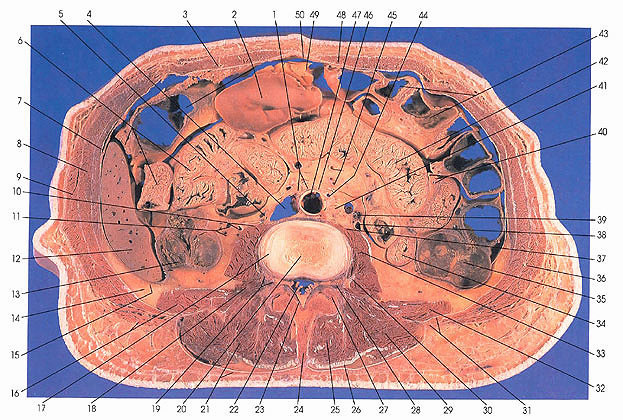
Anatomy Atlases Atlas Of Human Anatomy In Cross Section Section 5 Lower Thorax Lungs And Abdomen from www.anatomyatlases.org This muscle forms the anterior and lateral abdominal wall. But with the use of smart technology, you can learn faster and master abdomen anatomy in no time! The above lines intersect and divide the abdomen into nine regions (clockwise from the top) Muscle performance in neck pain assessment and rehab of the deep. Muscle performance in neck pain online course: Two layers in abdomenfatty superficial layer (camper's fascia)deeper membranous layer (scarper's fascia). In order to find the right training and to perform the exercises properly, it is important to know what are the abdominal muscles. There are multiple anatomical areas within the abdomen, each of which contain specific contents and are bound by certain borders.
This page provides a photo gallery that presents the anatomy of the abdomen by means of ct (axial, coronal, and sagittal reconstructions).
Windham was previously a surgical oncologist in the sarcoma program of the h. Level of l5, near transtubercular plane anatomy ileum, rectus abdominis muscle, ileocecal junction, cecum, internal abdominal oblique muscle, external abdominal oblique muscle, psoas major muscle, iliacus muscle, body of l5 vertebra. The abdomen (colloquially called the stomach, belly, tummy or midriff) is the part of the body between the thorax (chest) and pelvis, in humans and in other vertebrates. A good amount of area is covered by the abdominal wall. The above lines intersect and divide the abdomen into nine regions (clockwise from the top) The problem is that the basic crunch is. Simple, easy notes for quick revision of important questions. 6 write the origin, insertion and nerve supply of muscles of anterior abdominal wall. Divided into 9 regions by two vertical and two horizontal imaginary planes. Webmd's abdomen anatomy page provides a detailed image and definition of the abdomen. Having visible abs is a byproduct of nutrition, exercise, and overall caloric expenditure. Like , comment , share , subscribe #tcml #charsi whatsapp : These general diagrams show the digestive system, with the major human anatomical structures labeled (mouth, tongue, oral cavity, teeth, buccal glands, throat, pharynx, oesophagus, stomach, small intestine, large.
Share :
Post a Comment
for "Abdominal Anatomy / Abdominal Wall Overview Anatomy Lecturio Youtube"

Post a Comment for "Abdominal Anatomy / Abdominal Wall Overview Anatomy Lecturio Youtube"